
Retinal migraine is an extremely rare condition involving repeated, fully reversible visual changes in one eye, usually with a migraine headache. It is a diagnosis of exclusion: clinicians must first rule out more common and potentially dangerous causes of temporary monocular vision loss. It is not a safe catch-all name for flashes, blind spots, or “ocular migraine.”
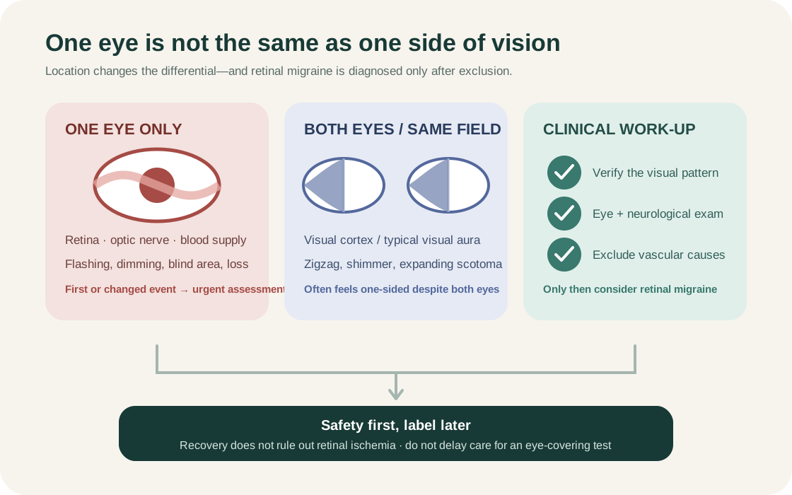
The distinction matters because typical migraine visual aura begins in the brain and affects the visual field of both eyes, even when the disturbance seems stronger on one side. A truly one-eye symptom can arise from the retina, optic nerve, or blood supply to the eye and deserves prompt assessment.
Get urgent medical assessment for a new visual change that appears to affect one eye. Call emergency services for sudden inability to see, a curtain or shadow over vision, visual symptoms with weakness, numbness, speech trouble, confusion, fainting, or a sudden severe headache. Do not drive or delay care to test which eye is involved.
What is retinal migraine?
The International Classification of Headache Disorders (ICHD-3) describes retinal migraine as repeated attacks of monocular visual disturbance—such as scintillations, a blind area, or blindness—associated with migraine headache. “Monocular” means the phenomenon is present in one eye, not merely on one side of the scene seen by both eyes.
Retinal migraine is far less common than migraine with visual aura. Many reported cases do not satisfy formal criteria or do not adequately exclude other causes. That is why a clinician should not infer the diagnosis from the word “migraine,” a family history, or recovery of sight alone.
Retinal migraine symptoms
Reported retinal migraine symptoms can be positive, meaning something is added to vision, or negative, meaning part of vision is missing.
- Positive phenomena can include flashing, sparkling, shimmering, or scintillating light.
- Negative phenomena can include a dim area, blind spot (scotoma), narrowing, or temporary loss of vision.
- The change is fully reversible under the formal definition.
- It develops gradually over at least five minutes, lasts 5–60 minutes, and/or is accompanied or followed within 60 minutes by headache; at least two of those timing features are required.
Those features describe the classified disorder, but none is a home safety test. Ischemic and other eye conditions can also be temporary. Persistent loss is not retinal migraine under the criteria and needs emergency assessment.
Retinal migraine vs. visual aura
Typical visual aura originates in the visual-processing regions of the brain. It often creates a zigzag, shimmering arc, expanding blind spot, or distortion that occupies the same side of the visual field in both eyes. Because attention is drawn to one side, people commonly remember it as happening in one eye.
Retinal migraine is different: the disturbance must be confirmed as monocular. In practice, that can be difficult after an event. ICHD-3 accepts confirmation during an attack by clinical visual-field examination or by a patient drawing a monocular field defect after clear instruction.
If a familiar, previously assessed event occurs and a clinician has given safe instructions, observations may help. But during a first or changed event, do not repeatedly cover the eyes, stare at bright patterns, or postpone urgent assessment. Read the broader migraine aura guide for sensory, language, and visual aura patterns.
Why “ocular migraine” causes confusion
“Ocular migraine” is not a precise diagnosis. People use it for both common cortical visual aura and rare retinal migraine. The two have different anatomical locations and different safety implications.
Mayo Clinic notes that visual changes in only one eye usually are not retinal migraine and may need treatment right away. When talking with a clinician, describe exactly what was seen and how it changed instead of relying on “ocular migraine.” Our ocular migraine terminology guide explains the naming problem in more detail.
Formal retinal migraine diagnostic criteria
ICHD-3 requires attacks that satisfy migraine-with-aura criteria plus all of the following:
- Fully reversible positive and/or negative visual phenomena in one eye.
- Monocular involvement confirmed by examination during an attack or an instructed drawing.
- At least two of three features: gradual spread over five minutes or more, duration of 5–60 minutes, and headache during the symptom or within 60 minutes afterward.
- No better ICHD-3 diagnosis.
- Exclusion of other causes of amaurosis fugax (temporary visual loss).
Classification criteria help trained clinicians describe a recurring pattern. They are not a checklist for self-diagnosis, and a first event cannot establish the repeated-attack requirement.
What else can cause one-eye vision loss?
Transient monocular vision loss has a broad differential. It can arise from reduced blood flow to the retina or optic nerve, embolic disease, carotid disease, inflammation such as giant cell arteritis, retinal or vitreous problems, intermittent angle-closure glaucoma, optic-disc disorders, or other neurological and ophthalmic conditions.
The history gives important clues but cannot safely exclude these causes online. A descending curtain, a new shower of floaters and flashes, painful red eye, jaw pain or scalp tenderness in an older adult, or persistent deficit changes the urgency. A painless symptom can still be serious.
AAO EyeWiki describes ischemic amaurosis fugax as a form of transient ischemic attack and a possible warning of stroke. Recovery therefore does not prove that an episode was harmless.
| Cause | Typical features | Key difference from retinal migraine |
|---|---|---|
| Migraine visual aura | Binocular zigzag/shimmer in same visual field of both eyes; 20–30 min; no eye pain | Binocular, not monocular; cortical, not retinal origin |
| Amaurosis fugax (TIA/embolic) | Monocular dim or curtain; seconds to minutes; may be painless; carotid or cardiac source | Vascular emergency; often abrupt and painless; not migraine-related |
| Retinal detachment | Flashes, floaters, then shadow or curtain; may be persistent | Progressive and structural; does not resolve fully without treatment |
| Acute angle-closure glaucoma | Painful red eye; halos around lights; nausea; raised IOP | Pain and redness are hallmarks; requires urgent pressure-lowering |
| Giant cell arteritis (GCA) | Age >50; jaw claudication; scalp tenderness; raised ESR/CRP; sudden vision loss | Systemic inflammatory markers; optic nerve ischemia risk |
| Retinal vein/artery occlusion | Sudden, often painless; visual field loss; fundus changes on examination | Structural, non-reversible; detected on dilated examination |
| Optic neuritis | Central blur or dimness; color desaturation; pain on eye movement; may be demyelinating | Pain on movement; colour vision loss; no aura timing |
This table summarises key distinguishing points; it is not a diagnostic tool. A clinician performs the examination and targeted investigation needed to differentiate these conditions.
When to seek urgent or emergency care
Use emergency services for:
- sudden complete or major vision loss in either eye;
- a curtain, veil, or shadow that does not clear;
- visual change with weakness, numbness, facial droop, speech difficulty, severe imbalance, confusion, or loss of consciousness;
- a sudden “worst” or thunderclap headache;
- severe eye pain, red eye, halos, nausea, and reduced vision; or
- visual loss after significant head or eye injury.
Seek prompt same-day professional advice for a first one-eye episode, a newly changed pattern, symptoms outside the usual 5–60-minute aura window, new floaters/flashes, or events beginning after age 50. Pregnancy/postpartum status, vascular disease, clotting history, and new medicines also belong in the urgent history. Follow local emergency guidance; do not drive yourself while vision is impaired.
How retinal migraine is diagnosed
Evaluation begins with a precise timeline and examination, often involving ophthalmology and sometimes neurology or stroke services. The clinician may assess visual acuity and fields, pupils, eye movements, the retina and optic nerve, blood pressure, neurological findings, and vascular risk.
Tests depend on age, examination, duration, and suspected alternatives. They can include dilated retinal examination, optical coherence tomography, visual-field testing, retinal imaging, blood tests for inflammatory or metabolic causes, electrocardiography, carotid or cardiac assessment, and brain/vascular imaging. A normal test after symptoms resolve does not automatically establish migraine; the whole clinical pattern and exclusion work matter.
Bring an up-to-date medication list, including hormonal contraception, over-the-counter products, and recent changes. Mention smoking or nicotine use, pregnancy/postpartum status, cardiovascular history, eye disease, and relatives with early vascular disease.
What causes retinal migraine?
The mechanism remains uncertain. Retinal or ophthalmic-artery vasospasm has often been proposed, but the evidence is limited and the label has been applied inconsistently. Reviews emphasize that many apparent cases may instead be cortical aura or another cause of transient monocular vision loss.
This uncertainty matters for treatment claims. A trigger does not prove retinal vasospasm, and eliminating a food or screen exposure cannot exclude vascular or eye disease. The defensible conclusion is narrower: recurrent visual episodes deserve accurate localization and a diagnosis based on exclusion, not speculation.
Retinal migraine treatment
There are no large, high-quality treatment trials specific to retinal migraine. After urgent causes have been excluded and a specialist has made the diagnosis, management is individualized around attack frequency, disability, headache phenotype, vascular context, and medicine safety.
For infrequent short episodes, a clinician may prioritize observation, safety planning, and treatment of the associated headache. For recurrent or disabling attacks, migraine-preventive medicines may be considered. Published reports mention options such as calcium-channel blockers, but case reports and small series do not establish a universal first choice.
Some reviews advise caution with vasoconstrictive medicines such as triptans or ergots when transient monocular loss is suspected, because the retinal mechanism is uncertain and ischemia must be excluded. Do not start, stop, or borrow migraine medicine based on an online label. A specialist should decide whether an acute or preventive treatment fits the confirmed diagnosis and cardiovascular profile.
Risk-factor and trigger management
General migraine routines—regular sleep, meals, hydration, activity, and consistent caffeine—may reduce overall attack variability for some people, but they do not treat a retinal emergency. Avoid presenting every event as a reaction to screens, stress, dehydration, or a particular food.
Clinicians may also address modifiable vascular risk: blood pressure, diabetes, cholesterol, smoking/nicotine, and medication or hormonal factors. This is risk-informed care, not proof that those factors caused the episode. If smoking and estrogen-containing contraception coexist with migraine aura or unexplained visual loss, discuss the combination with a qualified clinician rather than changing prescribed contraception without a plan.
What to do during a visual event
- Stop driving, cycling, using machinery, or climbing.
- Sit or lie somewhere safe and note the start time.
- Use the urgent-care plan previously given by a clinician; for a first or changed one-eye event, obtain urgent assessment.
- Do not test vision with bright light, wait for a tracker timer to finish, or take unprescribed aspirin or someone else’s migraine medicine.
- If safe, ask another person to stay nearby and help record what you describe in real time.
Safety comes before documentation. If emergency signs are present, call first and record later.
How to track a monocular visual episode
After care is arranged, record details that help reconstruct the event:
- exact start, peak, end, and whether vision fully returned;
- the person’s own description: flashing, blur, dark patch, curtain, missing area, or distortion;
- whether one eye was clinically confirmed, merely suspected, or unknown;
- how the shape moved or spread;
- headache timing, side, severity, nausea, and light/sound sensitivity;
- eye pain, redness, floaters, weakness, numbness, speech or balance changes;
- activity, posture, injury, recent illness, medicines, and menstrual/pregnancy context; and
- whether this was first-ever, familiar, or different from previous events.
A freehand drawing made after clear clinical instruction can sometimes be more useful than a long label. Keep raw observations separate from interpretations such as “retinal migraine.” Calmraine can record a timeline, but it cannot determine which eye or tissue produced a symptom.
Frequently asked questions
Is retinal migraine dangerous?
The classified episodes are reversible, but retinal migraine is a diagnosis of exclusion and rare permanent visual loss has been reported. More importantly, other causes of one-eye visual loss can threaten vision or signal vascular disease. New symptoms require prompt assessment.
Can retinal migraine affect both eyes?
No. By definition, retinal migraine is monocular. A visual pattern affecting corresponding parts of both eyes is more consistent with cortical visual aura, though a clinician must interpret the history.
How long does retinal migraine last?
ICHD-3 criteria use a 5–60-minute window as one of three timing features, with at least two required. Shorter, longer, or persistent visual changes need medical assessment rather than automatic reclassification.
Can retinal migraine happen without headache?
Cases without headache have been reported, but ICHD-3 notes that migraine as the cause cannot then be confidently established. One-eye visual loss without headache still needs investigation.
Is there a test for retinal migraine?
There is no single confirmatory scan or blood test. Diagnosis depends on a verified monocular recurring pattern, examination, and appropriate tests excluding retinal, optic-nerve, inflammatory, and vascular alternatives.
The practical takeaway
Retinal migraine means repeated, reversible visual phenomena truly confined to one eye and associated with migraine headache after other causes have been excluded. It is not the same as the far more common visual aura affecting both eyes, and it should not be inferred from the vague term “ocular migraine.” Treat a first, changed, or persistent one-eye visual symptom as a reason for urgent assessment. Once safety is addressed, a precise timeline, raw visual description, examination, and targeted work-up are more useful than any self-applied label.
For adjacent patterns, see silent migraine, photophobia and light sensitivity, and the medical disclaimer.
Calmraine provides educational information and cannot diagnose visual symptoms or retinal migraine. Read the medical disclaimer.
Sources
- 1.2.4 Retinal migraineInternational Headache Society · Accessed
- 1.2 Migraine with auraInternational Headache Society · Accessed
- Ocular migraine: When to seek helpMayo Clinic · Accessed
- Understanding ocular migraineAmerican Migraine Foundation · Accessed
- Vision lossNHS · Accessed
- Amaurosis Fugax (Transient Vision Loss)American Academy of Ophthalmology EyeWiki · Accessed
- Current Perspective on Retinal MigraineVision / PubMed Central · Accessed